Slot Scanning Mammography
- Slot Scanning Mammography Software
- Slot Scanning Mammography Imaging
- Slot Scanning Mammography Services
A 3D mammogram looks one page at a time,” Dr. Attai, MD, an assistant clinical professor in the department of surgery at David Geffen School of Medicine at the University of California.
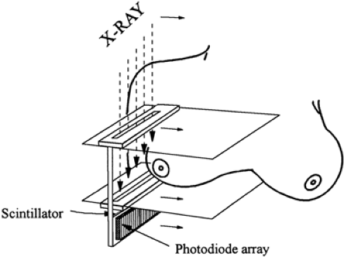
- Full field digital mammography systems cannot employ conventional AEC methods because digital receptors fully absorb the x‐ray beam. In this paper we describe an AEC procedure for slot scanning mammography. With slot scanning detectors, our approach uses a fast low‐resolution and low‐exposure prescan to acquire an image of the breast.
- PURPOSE:To evaluate a comprehensive array of scatter cleanup techniques in mammography by using a consistent methodology.
- Screening Mammography. Digital mammography is the only screening method that is consistently proven to reduce breast cancer deaths. Current guidelines from the U.S. Department of Health and Human Services, American Cancer Society, American Medical Association and American College of Radiology recommend screening mammography every year for women, beginning at age 40.
Slot Scanning Mammography Software
Slot scanning versus antiscatter grid in digital mammography: comparison of low-contrast performance using contrast-detail measurement
Abstract
Slot scanning imaging techniques allow for effective scatter rejection without attenuating primary x-rays. The use of these techniques should generate better image quality for the same mean glandular dose (MGD) or a similar image quality for a lower MGD as compared to imaging techniques using an anti-scatter grid. In this study, we compared a slot scanning digital mammography system (SenoScan, Fisher Imaging Systems, Denver, CO) to a full-field digital mammography (FFDM) system used in conjunction with a 5:1 anti-scatter grid (SenoGraphe 2000D, General Electric Medical Systems, Milwaukee, WI). Images of a contrast-detail phantom (University Hospital Nijmegen, The Netherlands) were reviewed to measure the contrast-detail curves for both systems. These curves were measured at 100%, 71%, 49% and 33% of the reference mean glandular dose (MGD), as determined by photo-timing, for the Fisher system and 100% for the GE system. Soft-copy reading was performed on review workstations provided by the manufacturers. The correct observation ratios (CORs) were also computed and used to compare the performance of the two systems. The results showed that, based on the contrast-detail curves, the performance of the Fisher images, acquired at 100% and 71% of the reference MGD, was comparable to the GE images at 100% of the reference MGD. The CORs for Fisher images were 0.463 and 0.444 at 100% and 71% of the reference MGD, respectively, compared to 0.453 for the GE images at 100% of the reference MGD.
Slot Scanning Mammography Imaging

Slot Scanning Mammography Services
- Publication:
- Pub Date:
- May 2004
- DOI:
- 10.1117/12.535986
- Bibcode:
- 2004SPIE.5368..734L